NEWS
IROAST Researchers - Prof. Takumi Higaki
English Japanese
Newly opened academic field that quantitatively captures plant cells using imaging

Professor Takumi Higaki
IROAST International Joint Research Faculty Member
Faculty of Advanced Science and Technology
(Period at IROAST: from August 2017 to September 2021)
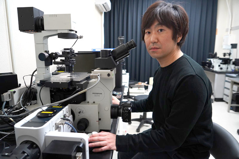
At Kumamoto University, a professor has opened up a new academic field that takes quantitative measurements of items using imaging and a computer, where previously they could only be qualitatively analyzed through a microscope. That person is Professor Takumi Higaki, who obtained tenure in October 2021.
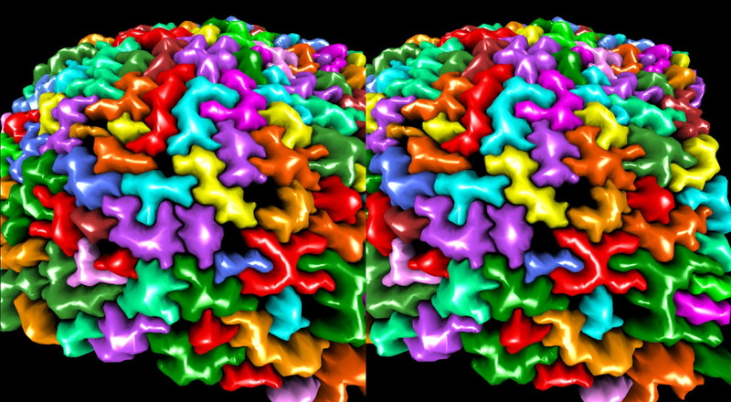
■ Viewing plant cells in a world other than 2D using equipment such as VR goggles and 3D scanners
Q: Please tell us about the content of your current research.
Higaki: We conduct research such as observing structures inside plant cells using a microscope, such as the cytoskeleton and organelles, which change from moment to moment, and analyzing the shape and movement of cell structures using a computer. With the commonly used visual observation through a microscope, the way the cells were viewed and interpreted differed from person to person, and the results could even differ for the same person, depending on how they were feeling physically and their mood. However, when cells are measured using a computer, you can expect to obtain the same results regardless of when the cells are measured and by whom. This is called reproducibility, and it is very important in science. There are also elements that are not noticed through visual observation alone, but become clear with computer-based analysis. Thus, we are conducting research to improve the observation ability of humans with imaging analysis using computers.
However, careful observation of an item by humans is considered to be just as important as analysis using a computer. Until now, the main way of viewing cells was to look directly through a microscope lens or to observe a flat display of a captured image, but recently it has become possible to observe a cell using VR in a way that seems like you have entered the cell. We are conducting research on the usefulness of this new observation method.

Q: Have you encountered research that has been especially rewarding from the time when you first joined IROAST to now?
Higaki: My research until now has been at the cellular level, mainly using microscopes, but during my time at IROAST I obtained information on the 3-dimensional shape at the individual plant level using a single-lens reflex camera in collaboration with a professor from the Faculty of Engineering. We then successfully developed a system to quantitatively evaluate plant characteristics such as the angle of branches. It is like a high-precision 3D scanner for plants. This causes almost no damage to the plant, so it can be kept as a digital archive of the plant’s growth process.
Successfully expanding the research subject from the cellular level to the individual level using the common technology of imaging analysis is one of the events that has made me very happy since joining IROAST.

■ Collaborative research developed through exchanges between an engineering professor and IRCMS. The environment of IROAST that allows you to concentrate on your research is very appealing.
Q: What brought you to IROAST?
Higaki: I was a fixed-term faculty member at a university in the Kanto region, but the professor in my laboratory was about to retire, so I thought I’d strike out on my own. Then I saw the open call for IROAST participants. I am originally from Oita, so I was already familiar with Kumamoto.
Q: What is the appeal of IROAST?
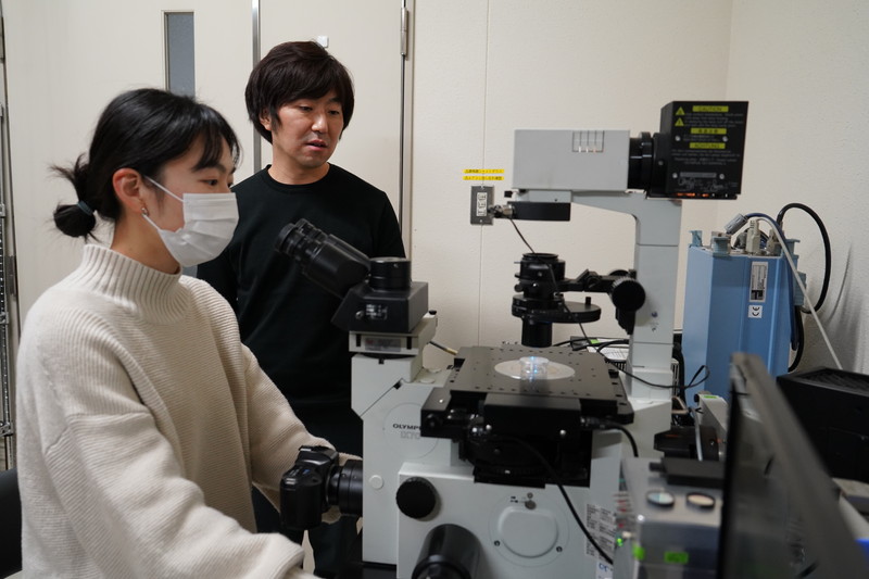
Higaki: There are many researchers from the Faculty of Science and the Faculty of Engineering at IROAST, which leads to proactive exchange between different fields of study. For example, I was contacted by Professor Rude Lee, who was on the same IROAST tenure track (currently at the Institute of Industrial Nanomaterials), and we conducted joint research within IROAST. Professor Lee is an expert in nanomedicine, and I assisted in the evaluation of a lesion detection probe she developed. I am in the Faculty of Science, but I feel that IROAST gave me the opportunity to exchange ideas with a professor from the Faculty of Engineering.
IROAST also conducts proactive exchange with IRCMS (Kumamoto University International Research Center for Medical Sciences), and I am also continuing collaborative research with professors conducting basic medical research at IRCMS. Bringing together different areas of technical expertise in this kind of collaborative research enables significant research that could not be achieved by an individual researcher.
Q: How good is the tenure track system?
Higaki: It provides an environment that allows you to concentrate on your research. University faculty staff have many other duties outside their own research, such as teaching and management. Thus, in that respect, tenure track staff can concentrate on their own research.
On the other hand, there were many opportunities to learn about university education and management during the tenure track period, which enabled a relatively smooth transition after obtaining tenure.

■ I want to train young researchers to be capable in both biology and image analysis
Q: What is your future outlook?
Higaki: One of the characteristics of images is that they are able to express a broad range of spatial scales handled in biological science, from molecules to ecology. So, I want to conduct research beyond the spatial scale through analysis of images at a broad variety of magnifications. For example, at the moment I am conducting research that considers the relationship between the behavior of individual plant cells and the shaping of organs, which are collections of cells. This is research connecting the different spatial scales of cells and organs, but in the future, I think it may also tie in with more detailed structural analysis using special microscopes and conversely large-scale analysis using equipment like drones and satellite images. Either way, for me, I want to understand living organisms through images.
From now on, I also want to concentrate on educating the younger generation. There are still very few people capable in both biological science and image analysis, so I want to convey how important and interesting image analysis is to students majoring in biological science. Conversely, I also think it is important to convey the appeal of biological science to engineering students. I want to contribute to the development of this field of research as much as possible by fostering students and young researchers.
Q: What is your message to young researchers?
Higaki: In addition to IROAST, Kumamoto University has many centers conducting outstanding world-class research, including IRCMS, the Institute of Molecular Embryology and Genetics, the Magnesium Research Center, and the Institute of Industrial Nanomaterials. Therefore, not only are individual researchers conducting high levels of research, there is also a strong tradition of interaction among young researchers.
Kumamoto is also a lovely place to live. After coming to Kumamoto, I found my commute and living environment became very pleasant. I would also recommend Kumamoto as a great place to raise children, so I would definitely recommend research at Kumamoto University as a good option.
Reference links
- Imaging Biology, Faculty of Science, Kumamoto University | Higaki Laboratory

