- HOME
- RESEARCH
- Research Units
- Quantification of Three Dimensional Vascular Network
RESEARCH
Quantification of Three Dimensional Vascular Network
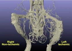
Blood vessels change their morphology according to various physiological and pathophysiological conditions. In the field of vascular biology, many biological mechanisms are hypothesized, however, it is very difficult task to quantify the three dimensional vascular architecture.
Our collaboration has already established the image processing method of vascular network (Arima et al., Journal of American Heart Association, 2018). The aim of our team is to establish the unique quantification method of three dimensional data.
Figure: 3D image of murine bone and vascular network in lower leg.

Unit members
-
 Yuichiro ARIMA
Associate Professor, International Research Center for Medical Sciences (IRCMS), Kumamoto University/X-Earth Center, Faculty of Engineering, Kumamoto University
Japan -
 Unit coordinatorToshifumi MUKUNOKI
Professor, Faculty of Advanced Science and Technology, Kumamoto University/X-Earth Center, Faculty of Engineering, Kumamoto University
Japan -
 Patrice DELMAS Website
Associate Professor
Department of Computer Science, The University of Auckland
*IROAST Visiting ProfessorNew Zealand
Achievements
Publications
Arima Y, Mukunoki T, et. al., Sample Preparation for Computed Tomography-based Three-dimensional Visualization of Murine Hind-limb Vessels. J Vis Exp. 2021
Grants
(Application)
Grants-in-Aid for Scientific Research-KAKENHI- Challenging research 2022, “CT-based immunostaining”
Activities
Presentation
Yuichiro Arima, “Visualization of blood vessel microstructure by CT ~Efforts of medical-engineering collaboration~”, The 8th International Workshop on X-Ray CT Visualization for Socio-Cultural Engineering and Environmental Materials, 2021.
- Units of World-leading Researchers
-
- Development of Nano and Supramolecular Materials
- RNA Biology
- Plant Cell and Developmental Biology
- Nano-Organics and Nano-Hybrids
- Nano-medicine and Drug Delivery System
- Nano-medicine and Theranostics
- Multiscale Modeling of Soil and Rock Materials Using X-ray CT
- Quantification of Three Dimensional Vascular Network
- MicroCT-based Quantification of Fibrosis and Vascularization in Pancreatic Tumor
- Advanced Structural Materials
- Microstructure Analysis and Grain Boundary Engineering
- Structure and Dynamics of Materials Using Quantum Beams and Data-Driven Sciences
- Hydrological Environments
- Nano-materials for Energy Applications and Environmental Protection
- Units of Young Researchers
-
- Quantitative Bioimaging
- Development of Novel Therapeutic Strategy Using Iron Targeted Upconversion Nanoparticles for Parkinson's Disease
- Deep Learning for Hydrology
- Environmental Impacts of Ionic Solutes
- Study of first-generation objects in the universe with radio telescopes
- Plant Stem Cells and Regeneration
- Development of Microbially-Aided Carbon Sequestration Technology
- Advanced Biomedical Evaluation System
- Bio-inspired Functional Molecular System
- Nanomaterials Processing for Medical, Cosmetic, and Environmental Applications
- Ferroelectric Photovoltaics
- Next-Generation Design of Structures


